Immunobiological Tests
1. Mixed lymphocyte reaction
Mixed lymphocyte reaction (MLR), also known as mixed lymphocyte culture, refers to the in vitro mixed culture of two unrelated individuals with normal lymphocytes. Due to the different HLA-II antigen D and antigen DP on the cell surface, they can stimulate each other's lymphocyte transformation and release lymphocyte factors such as IL-2. In the in vitro functional experimental program for antibody drug development, the activity of these antibody molecules is evaluated by detecting the amount of cytokines released in the MLR experiment.

In DC-T mixed lymphocyte reaction, T cells are activated and release cytokines IL-2 and IFN-Γ
2. T cell activation and proliferation experiments
The activation of T lymphocytes is the core of immune response. Inducing T cell activation, proliferation and differentiation into effector T cells requires dual signal stimulation. The most common antibodies used for in vitro activation of T cells are anti-CD3 antibodies and anti-CD28 antibodies. After T cells are activated, IL-2, IFN-Γ, etc. can be detected by FACS, ELISA and other methods to determine the state of T cell activation and proliferation.

FACS detection of aCD3/aCD28-induced mouse T cell activation and proliferation, including the increase of CFSE peak and cytokine IL-2 and IFN-Γ
 3. T cell killing function experiment
T cell-mediated cytotoxicity is a characteristic of cytotoxic T cells (CTLs). When sensitized T cells encounter the corresponding target cell antigens again, they can show destructive and lytic effects on the target cells. It is a commonly used indicator for evaluating the body's cellular immunity level. Clinically, it is especially used to measure the ability of CTLs in tumor patients to kill tumor cells, and is often used as one of the indicators for judging prognosis and observing therapeutic effects.
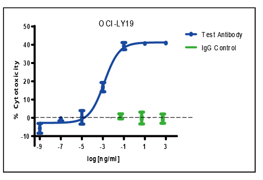
The antibody to be tested is a CD3/CD19 bispecific antibody; the effector cells are purified CD3+ T cells; the target cells are OCI-LY-19; the E:T ratio is 10:1; the co-incubation time is 4 hours;
4. Antibody-dependent cellular cytotoxicity assay ADCC
IgG antibodies can mediate these cells to exert ADCC effects, among which NK cells are the main cells that can exert ADCC effects. During the process of antibody-mediated ADCC, antibodies can only specifically bind to the corresponding antigen epitopes on target cells, while effector cells such as NK cells can kill any target cells that have bound to antibodies. Therefore, the binding of antibodies to antigens on target cells is specific, and the killing effect of NK cells on target cells is non-specific.

5. Complement-dependent cell killing assay CDC
Complement dependent cytotoxicity (CDC) refers to the cytotoxic effect of complement, that is, the classical complement pathway is activated by the binding of specific antibodies to the corresponding antigens on the cell membrane surface to form a complex. The membrane attack complex formed has a lytic effect on the target cells.

6. Flow cytometry (FACS)


